


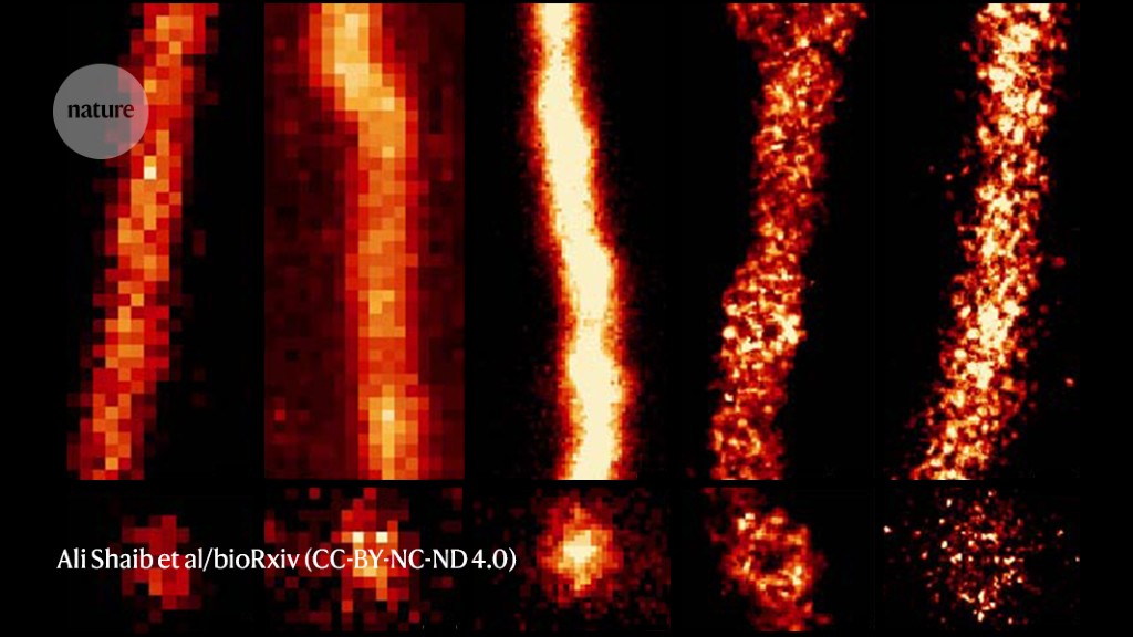
The protein tubulin imaged using existing super-resolution and expansion microscopy methods (panels 1–3) and the ONE microscopy technique (panels 4 and 5).Credit: Ali Shaib et al/bioRxiv(CC-BY-NC-ND 4.0)
When Ali Shaib was doing his master’s degree at the Lebanese University in Beirut, he spent several weeks on a waiting list and visited a different campus to take a few images on a costly microscope, something scholars in richer countries took for granted.
Now, Shaib, a nanoscale specialist at the University Medical Center Göttingen in Germany, and his colleagues have developed a method for ordinary light microscopes that they hope will demolish such barriers.
Revolutionary microscopy technique sees individual atoms for first time
The technique1 — which has recorded jaw-dropping images of individual proteins and never-before-seen structures in cells — offers a level of detail that eclipses even that of multi-million-dollar ‘super-resolution’ microscopes.
“There should be some form of democracy in microscopy,” says Silvio Rizzoli, a nanoscale specialist also at the University Medical Center Göttingen who has pioneered the technique, dubbed ONE microscopy, with Shaib. “It’s high resolution for the many, not the few rich labs.”
At the nanoscale
The power of conventional light microscopes is limited by the laws of optics, which mean that objects smaller than about 200 nanometres are a blur. Researchers have developed physics-beating super-resolution methods that, Rizzoli says, can bring this limit down to around 10 nm. The approach, which earned the 2014 Nobel Prize in Chemistry, uses optical tricks to pinpoint fluorescent molecules attached to proteins.
In 2015, researchers came up with another way to evade optical limits. A team led by Edward Boyden, a neuroengineer at the Massachusetts Institute of Technology in Cambridge, showed2 that inflating tissue — using an absorbent compound in nappies — moves cellular objects away from each other. This technique, called expansion microscopy, led to leaps in microscope resolution and can resolve structures of around 20 nm.
Shaib and Rizzoli’s technique — described in a study posted to the bioRxiv preprint server last month — melds the two approaches to achieve resolutions below 1 nm. That is sharp enough to reveal the shape of individual proteins, which are typically imaged in finer detail using much more expensive structural-biology methods such as cryo-electron microscopy (cryo-EM) or X-ray crystallography.
Expansion microscopy’s simplicity is part of its appeal, says Boyden, who estimates that more than 1,000 laboratories have adopted the technique. Samples are treated with chemicals that anchor their proteins to a polymer that, with the addition of water, swells to 1,000 times its size, moving the molecules apart. ONE (short for one-step nanoscale-expansion) microscopy uses heat or enzymes to also break the proteins apart, so that individual fragments are stretched in different directions during expansion.
This conventional confocal microscope can achieve nanoscale resolutions using the ONE microscopy technique.Credit: Ali Shaib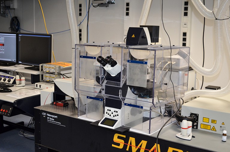
The researchers have used their approach to record pictures of a neural molecule, the GABAA receptor, that closely resemble much-higher-resolution cryo-EM and X-ray crystallography maps of the protein. They also captured the outlines of a bulky protein called otoferlin, for which the structure hasn’t been determined and that helps to convey audio signals in the brain. The shape resembles a structural prediction made by the AlphaFold deep-learning network.
The method cannot match the resolution of cryo-EM, which can reveal near-atomic-level details smaller than 0.2 nm in some cases. But cryo-EM can be finnicky and expensive. By contrast, ONE microscopy could offer a quick and easy way to obtain structural insights into just about any molecule, says Rizzoli. “You can look at any protein, and you can get resolution you couldn’t dream about.”
Increased accessibility
Rizzoli, who is originally from Hungary, says that part of the motivation for developing the technique was to broaden the accessibility of cutting-edge light microscopy. The ONE-microscopy method is straightforward to apply and works with now-antiquated fluorescent microscopes from the 1990s.
Salma Tammam, a pharmaceutical technologist at the German University in Cairo, is planning to send a PhD student to Göttingen to learn the technique this summer. Her lab studies how nanoparticles move about in cells. They would like to see the fine details of the particles and their cargo. But like many researchers in low and middle-income countries, they do not have access to expensive super-resolution microscopes. “This brings us resolution in an affordable manner,” she says.
Revolutionary cryo-EM is taking over structural biology
Broadening the reach of super-resolution microscopy is also important for scientists at well-funded institutions, says Noa Lipstein, a synapse biologist at Leibniz Center for Molecular Pharmacology in Berlin. She has access to a well-established super-resolution technique called stimulated emission depletion microscopy. But she recently started an independent group and has chosen to apply ONE microscopy to their research into the fine details of neural synapses.
“It’s allowed me independence, because I don’t have to rely on connections to big shots with heavy machines,” Lipstein says. “This I can do in my own lab and my own bench.”
Lipstein hasn’t pushed the technique to its limits, but she’s already getting glimpses of new biology. “It’s almost a given that we are going to see new things. We already see them, but we don’t know what they are,” she says.
More News
How artificial intelligence is helping Ghana plan for a renewable energy future
Editorial Expression of Concern: Leptin stimulates fatty-acid oxidation by activating AMP-activated protein kinase – Nature
Quantum control of a cat qubit with bit-flip times exceeding ten seconds – Nature