


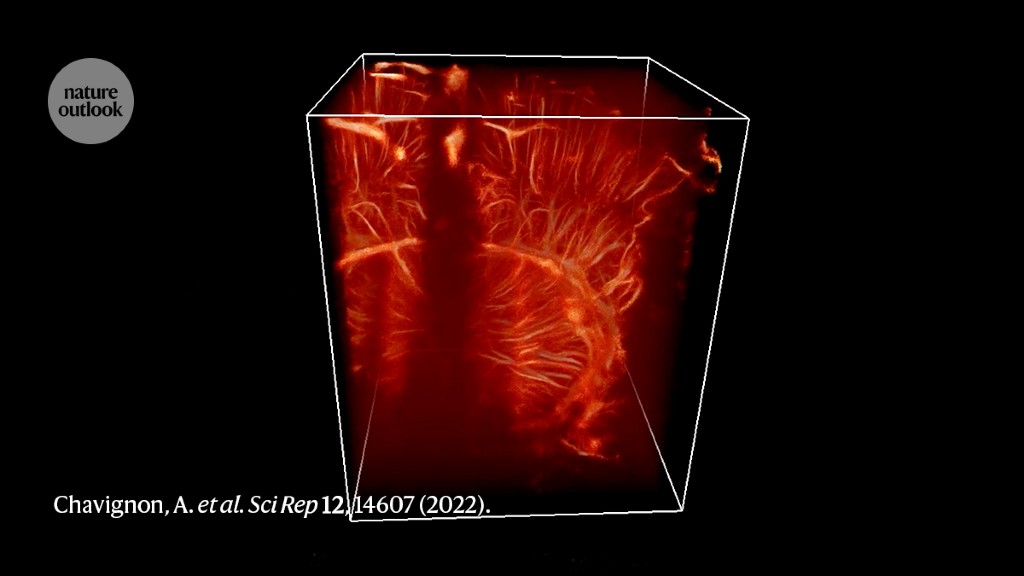
Ultrasound localization microscopy generates clearer images than conventional techniques.Credit: Chavignon, A. et al. Sci. Rep. 12, 14607 (2022).
Resolve Stroke in Paris spun off from Sorbonne University, Paris, in 2022.
Minutes matter when responding to a stroke. Delays in treatment can exacerbate damage to the brain and reduce the odds of recovery. Evidence suggests that surgery to remove any blood clots responsible for the attack must be done within seven hours of the start of a stroke1. Magnetic resonance imaging (MRI) or computed tomography (CT) are typically used to guide physicians to the clots, but these are generally done in hospital. Getting the person there, however, can delay stroke diagnosis, which usually requires a few hours.
Read more about The Spinoff Prize
Paris-based Resolve Stroke — a finalist for The Spinoff Prize 2023 — aims to boost the precision of and to accelerate stroke diagnosis with a simple, lightweight device at a hospital bedside or even in an ambulance. Its platform uses an imaging modality known as ultrasound localization microscopy (ULM). The technique was developed2 by Resolve Stroke’s co-founder and scientific adviser Olivier Couture in 2011, to generate micrometre-resolution, 3D images of blood flow through the labyrinth of blood vessels that traverse the brain. “What we are doing is basically filling the gap between microscopy, which is very local and very precise, and MRI or CT, which are more macroscopic imaging,” says Couture’s former PhD student Vincent Hingot, who co-founded Resolve Stroke and serves as its chief technology officer.
Conventional ultrasound uses a handheld probe to transmit high-frequency soundwaves into the body. As these waves pass through tissues with different material properties, they produce reflected echoes that are received by the probe and reconstructed into images. But these images typically have poor detail, with peak resolution in the order of hundreds of micrometres — sufficient to visualize only the thickest blood vessels.
Tracking bubbles
While working as director of research at the French National Centre for Scientific Research (CNRS), at Sorbonne University, Paris, Couture worked out a way to sharpen the pictures collected with ultrasound. Many ultrasound imaging systems use gas-filled microbubbles as a contrast agent; these are injected into the bloodstream, where they reflect back the ultrasound to reveal the contours of blood vessels. But because it is difficult to resolve individual bubbles, the resulting images are blurry. To address this shortcoming, ULM uses an ultrafast probe that captures hundreds or thousands of frames per second, enabling the detection of individual microbubbles in the circulation with a resolution approaching 10 micrometres.
In seconds, an intravenously delivered dose of millions of microbubbles — a process that uses inert gas and is already cleared for use in people in the United States, Europe and Japan — will begin to reach the brain. ULM then makes it possible to track their transit through some of the narrowest vessels, revealing constrictions and obstructions that might signify a stroke in progress. “Our technology is basically pinpointing individual microbubbles and following them in the bloodstream and reconstructing an image of the vasculature by summing all of these trajectories,” says Hingot. This imaging process can cover sizeable 3D volumes encompassing more than half of the human brain in a matter of minutes — just a few seconds is required if the focus is on large-diameter veins and arteries rather than a more complete vascular map (see ‘Sensing a stroke’).
Credit: Alisdair MacDonald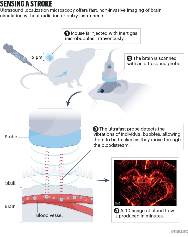
Detail without the bulk
Early adopters of ULM are enthusiastic about its potential. “The success here is unprecedented resolution combined with ease of use,” says Gianmarco Pinton, a biomedical engineer who works with ULM technology at the University of North Carolina at Chapel Hill. “You can just plop this device somewhere and all of a sudden you get extraordinary, exquisite detail of the vascular structure,” says Pinton, who is not connected to Resolve Stroke. This is in contrast to existing systems such as CT and MRI, which can deliver high-quality imaging but require bulky and costly equipment — and for CT, the use of radiation. Chief executive and co-founder of Resolve Stroke Aritz Zamacola says that its current instrument weighs 10 kilograms and could easily be transported by paramedics or wheeled into hospital rooms.
Part of Nature Outlook: The Spinoff Prize 2023
Resolve Stroke’s scientific team has reported several proof-of-concept animal studies, including a 2022 paper showing that it could detect and discriminate different forms of stroke using ULM to image through the skulls of live rats3. The team has also been working with sheep, which have skulls of similar thickness to humans. The start-up’s first-generation ULM probes should be capable of visualizing up to 60% of the human brain at a time. Zamacola says that it performs particularly well at the sites of larger blood vessels, which tend to give rise to more severe strokes and therefore require the most urgent intervention. Imaging at the capillary scale adds considerable time to the process.
Preparation for human testing is in the works. “We want to have a design freeze in the next year, and do a large clinical trial in 2024,” says Zamacola. The company’s goal is to enter the European clinical market in 2025.
Stroke is an ideal clinical test bed for ULM, but other opportunities await. Couture continues to run his academic laboratory at CNRS, and is pushing the imaging speed of ULM to achieve visualization of tiny capillaries that are currently difficult to resolve. These vessels play a central part in a variety of medical disorders, including neurodegenerative conditions such as Alzheimer’s disease. ULM’s capability is also relevant outside the brain; Couture is looking at blood filtration in the kidneys, for example, whereas Pinton has used ULM to study tumour circulation in animal models. “There are tons of different sites where the function of the organ is closely bound to the microcirculation,” says Couture.
Bringing an imaging technology to the clinical market is no small feat, and Resolve Stroke has a lot of work ahead. “Is ULM ready for full commercialization today?” says Pinton. “No — but I think the time is now for translation, and I think there are clear paths to commercialization for specific applications.” He does see some risk for the company in being the first to market — a disappointing debut for ULM in the clinic could lead to scepticism about the method. But he also thinks that Couture and his scientific co-founders have already demonstrated remarkable progress in their advancement of ULM’s capabilities, and he is confident that physicians will want to use it. “The clinicians I’ve talked to at conferences immediately see thousands of applications of this,” Pinton says. “They want this tool in their hands.”
More News
Author Correction: Bitter taste receptor activation by cholesterol and an intracellular tastant – Nature
Audio long read: How does ChatGPT ‘think’? Psychology and neuroscience crack open AI large language models
Ozempic keeps wowing: trial data show benefits for kidney disease